

Polish scientists are developing a robotic system for delivering drugs into the eye and performing biopsies. It is designed to assist ophthalmologists with procedures requiring exceptional precision, including gene therapies in which a drug must reach a precisely defined part of the retina. The technology being developed at the International Centre for Translational Eye Research is currently undergoing preclinical research. Ultimately, it is intended to support new treatments for eye diseases.
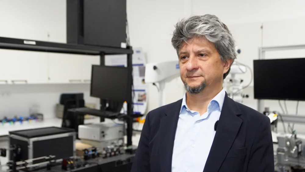
“We now have many new ways of treating the eye and vision that require support from the latest technologies, including delivering drugs very precisely to the place where they are meant to act. Eye surgery developed over many centuries, but it was only in the 20th century that it could advance very significantly. Such highly sophisticated robotic systems help limit damage to the eye when treatments are administered,” Professor Maciej Wojtkowski, head of the centre and of the International Research Agendas project, told Newseria.
The International Centre for Translational Eye Research (ICTER) project is being implemented under the International Research Agendas programme run by the Foundation for Polish Science and financed through European Funds for a Modern Economy (FENG).
A platform for new ophthalmic therapies
The project aims to build a platform supporting the development of new eye treatments. An automated drug delivery and biopsy system will be linked to imaging that identifies the position of an instrument inside the eye. The work also draws on expertise in gene therapy, single-cell genomics and bioinformatics.
“This is an entirely innovative project because such products are not yet on the market. Ophthalmologists very much need instruments that will help them test and introduce new treatments. These treatments are highly complex and can often be used only once—for example gene therapies, because our immune system learns to recognise the viruses that carry the genes, making it impossible to repeat the procedure. It must therefore be performed perfectly. Treatment effectiveness depends heavily on how the drug is administered,” Wojtkowski explained.
“I see the greatest potential for robotics and automation in supporting ophthalmologists and surgery in the elements that require greater precision, rather than necessarily those where the operator’s knowledge and experience are crucial. Certain final stages of a procedure, requiring a precise approach or movement along a defined axis, can be performed with a robot. But the ophthalmologist or operator decides, for example, on the angle of approach and can also stop the procedure while it is underway,” said Dr Karol Karnowski, a biophysicist at Nicolaus Copernicus University in Toruń and head of the ICTER project’s IDoc group.
Drug delivery into eye structures illustrates that division of responsibilities. The doctor chooses the insertion point and angle of the needle, while the robot can support precise movement along the selected axis. Sensors are also intended to help determine when the instrument reaches the correct position for administering the drug.
Precision measured in micrometres
“At present, the possibilities for administering drugs in gene therapy are very limited. It is a highly complex procedure in which a drug has to be delivered to a particular location on the retina with precision measured in individual micrometres—millionths of a metre. Such precision is currently achieved only by highly specialised operators. In future, we would like these procedures to be available everywhere. That is why this device is crucial,” Wojtkowski said.
The new technology is intended to be tested in work on optogenetic therapies for blindness, single-cell studies related to the development of uveal melanoma, and methods for the early detection of diabetic retinopathy.
ICTER’s robotics and automation research remains at the preclinical stage. Initial tests were performed on ex vivo models, meaning tissue outside a living organism. Researchers are preparing for in vivo studies in living organisms.
“The first tests use ex vivo models because they are more readily available. All the parameters are set, all the procedures tested, and then we can take further steps. Applications in humans are a completely different matter. These are not entirely scientific issues: they are more a subject for industry, which could take over the technologies and tools we develop. At a later stage, that would require compliance with all the regulations permitting use in humans,” Karnowski said.
Five research groups and PLN 30 million in funding
Five research groups with different specialisations are working on the project. Their cooperation is intended to enable the development and testing of the entire platform and its use in research into new ophthalmic therapies.
“Combining robotics with genetics, eye biology, bioinformatics and imaging gives us unique opportunities to develop a platform for introducing drugs and new therapies in ophthalmology. Without cooperation between groups from different fields, testing such a new robotic device and subsequently introducing it would not be possible. This combination of experience and knowledge from different disciplines is unique internationally and is offered only by our institute,” Wojtkowski said.
One component is imaging that tracks the position of an instrument relative to the structures of the eye. The IDoc team has previously worked on integrating a robotic manipulator with optical coherence tomography (OCT), which enables imaging of eye tissue.
The International Research Agendas project is funded through European Funds for a Modern Economy, with total financing of PLN 30 million. Researchers aim to advance the technology from TRL 4 to TRL 7: from laboratory validation to demonstrating a prototype in conditions approaching those of real use.
Source: Newseria.